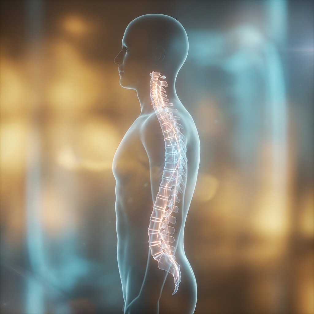
Cervical Postoperative Care Recovery Goals
Comprehensive postoperative care protocols for cervical disc replacement surgery including pain management, activity restrictions, rehabilitation programs, and long-term follow-up care to ensure optimal outcomes and successful recovery.
Early Mobilization
Rapid Recovery
24 Hours
Early mobilization within 24 hours post-surgery with structured rehabilitation protocols designed to optimize recovery while protecting the surgical site.
Success Rate
Proven Outcomes
95%
95% patient satisfaction rate with cervical disc replacement surgery based on comprehensive outcome measures including pain relief, function, and quality of life improvement.
Return to Work
Functional Recovery
6 Weeks
Most patients return to normal activities and work within 6 weeks following cervical disc replacement, significantly faster than traditional fusion surgery.
Immediate Postoperative ManagementThe Critical First Hours
The immediate postoperative period focuses on four primary clinical objectives: ensuring neurological stability, managing pain effectively through multimodal approaches, preventing common complications, and establishing the foundation for early mobilisation and recovery.
Outpatient or Short-Stay Setting
Most cervical disc replacement procedures are performed in outpatient or short-stay settings, with patients discharged within 24 hours postoperatively. This substantially shorter hospital stay compared to fusion surgery (traditionally 3–5 days) reflects the minimally traumatic anterior cervical approach and absence of bone graft healing requirements.
Neurological Stability
Continuous monitoring and immediate recognition of any changes from preoperative baseline
Effective Pain Management
Multimodal approach minimising opioid requirements whilst ensuring adequate comfort
Complication Prevention
Early identification and intervention for potential postoperative complications
Early Mobilisation
Establishing foundation for rapid functional recovery through early movement
Comprehensive Neurological Monitoring
Immediate neurological assessment occurs in the recovery room before patient awakening and continues at regular intervals throughout the first 24 hours, documenting any changes from preoperative baseline and identifying potential complications requiring urgent intervention.

Maintain or improve neurological status
The vast majority of patients maintain or improve preoperative neurological status. The excellent prognosis reflects that most patients experience immediate neurological improvement from decompression of previously compressed nerve roots or spinal cord.
Neurological Status Change Protocol
New or progressive neurological deficits require immediate action:
Early recognition and investigation is essential—early intervention significantly improves outcomes.
Multimodal Pain ManagementEvidence-Based Strategy
First 24 hours pain management utilises an evidence-based multimodal approach designed to provide adequate comfort whilst minimising opioid requirements and their associated side effects including nausea, constipation, sedation, and dependency risk.
Moderate Pain Expected
Some discomfort is normal and expected following surgery
Escalating Pain Warning
Progressively worsening pain may signal complications
Minimise Opioid Use
Excessive opioids interfere with mobilisation and increase complications
Medication Approach
Paracetamol (Acetaminophen)
Foundation of postoperative analgesia without gastrointestinal risks
Expected Pain Characteristics
Neck Incision Discomfort
Localised to anterior neck, mild to moderate intensity
Neck Stiffness
Common and mild to moderate
Referred Shoulder Pain
Reflects cervical nerve root irritation
Arm Pain Resolution
Should dramatically diminish or resolve entirely immediately after surgery
Escalating Pain
Pain that is progressively worsening or disproportionate to expected surgical trauma

Structured Opioid Weaning Protocol
~90% of patients weaned within 1–2 weeks
Maximum opioid dosing if needed; most patients transition to minimal doses by day 3–4
Reduce opioid frequency; use primarily for breakthrough pain
Minimal opioid use; primarily non-opioid analgesia
Discontinue opioids; continue non-opioid analgesics as needed
Swallowing and Airway ManagementDysphagia Protocols
Dysphagia (swallowing difficulty) is the most common postoperative symptom, affecting 15–30% of patients immediately following anterior cervical surgery due to soft tissue swelling, temporary pharyngeal muscle dysfunction, and normal inflammatory response.
Dysphagia Incidence
Monitoring Protocol
Speech pathology evaluation before first food or fluid intake, establishing baseline swallowing function and aspiration risk.
Diet Progression
Liquid Diet
Initiated first once swallowing assessment complete
Soft Foods
Progresses as swallowing improves
Regular Diet
Advanced as tolerated
Red Flag Symptoms Requiring Urgent Evaluation
Resolution Timeline
Some patients experience intermittent swallowing difficulty for months, but complete resolution occurs in >95% within 6–8 weeks.
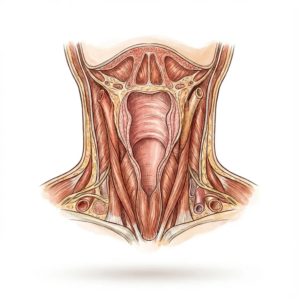
Anterior cervical anatomy: Delicate soft tissue structures affected during surgical approach require careful monitoring during recovery.
Early MobilisationThe Cervical Disc Replacement Advantage
Mobilisation beginning within hours represents a defining advantage of disc replacement compared to fusion surgery. Unlike fusion—which requires external immobilisation for 6–12 weeks to permit bone healing—disc replacement permits immediate mobilisation without external restriction.
Cervical Disc Replacement
- Immediate mobilisation within hours
- No external immobilisation required
- Motion preservation maintained
Traditional Fusion
- External immobilisation 6–12 weeks
- Restricted motion for bone healing
- Prolonged recovery timeline
Mobilisation Timeline and Progression
- Arm and leg movement in bed
- Active assistance to sitting on bed edge with support
- Ambulation with nursing or therapy assistance
- Short walks (10–20 feet) with support
- Independent or assisted ambulation
- Stairs with supervision
- Bathroom facility use
Physiological Benefits of Early Mobilisation
VTE Prevention
Muscle contraction during movement maintains venous return, substantially reducing blood clot risk
Pneumonia Prevention
Mobilisation enables lung expansion and respiratory secretion clearance
Deconditioning Prevention
Early movement preserves muscle function and cardiovascular fitness
Pain Reduction
Paradoxically, movement typically reduces pain through reduced stiffness and muscle tension
Psychological Benefit
Early mobility enhances confidence and mood; demonstrates functional recovery
Faster Recovery
Foundation for accelerated return to normal activities and work
Neck Movement Guidance
Although mobilisation is encouraged, patients should follow these guidelines to maintain normal motion patterns whilst respecting the healing surgical site:
Natural Self-Limitation
Most patients naturally self-limit extreme motions due to mild discomfort, minimising the need for additional restrictions. The goal is to maintain normal motion patterns whilst respecting the healing surgical site.

Discharge Criteria and Home ReadinessTransition to Recovery
Most patients are suitable for discharge within 24 hours following cervical disc replacement, provided that clearly defined medical and social criteria are satisfied.
Medical Stability Criteria for Discharge
Stable Vital Signs
Blood pressure, heart rate, respiratory rate, temperature maintained without support
Neurologically Stable
Stable or improved compared to preoperative baseline
Adequate Pain Control
Achievable with oral medications (no IV analgesia required)
Swallowing Function
Adequate for oral intake
No Concerning Signs
Fever, excessive swelling, wound complications absent
Independent Ambulation
Walking without significant assistance
Home Environment Readiness
Comprehensive Discharge Education
Before hospital discharge, patients and caregivers receive detailed written and verbal instruction:
Expected Symptoms: First Week at Home
Understanding normal recovery processes helps patients distinguish expected changes from concerning complications.
Neck Discomfort and Stiffness
Most pronounced days 2–3, gradually improving; worse with morning stiffness
ExpectedMild Anterior Neck Swelling
Typically peaks at 24–48 hours, gradually resolving over weeks
ExpectedReferred Shoulder Pain
Common; reflects nerve root irritation; typically resolves within days to one week
ExpectedFatigue
Marked fatigue is normal; most patients sleep 12–16 hours daily initially
ExpectedSleep Disturbance
Some experience sleeping difficulty due to position limitations; gradual improvement
ExpectedAppetite Changes
Mild reduction is common; usually brief duration
ExpectedWound Care and Incision Management
Incision Assessment
Daily observation for mild redness at incision borders (normal); spreading erythema, excessive warmth, swelling, or purulent discharge requires medical evaluation
Showering
Permitted within 48–72 hours with waterproof dressing protection; submersion (baths) deferred until fully healed (2–3 weeks)
Suture Removal
Typically 10–14 days if non-absorbable; many surgeons use absorbable sutures or skin adhesive
Scar Management
Initial scars gradually fade over months to years; sunscreen protection helps prevent darkening

Early Recovery PhaseActivity Progression and Work Return
Activity restrictions during the first 6 weeks aim to protect the anterior surgical site and healing implant-bone interface whilst avoiding excessive immobilisation that promotes stiffness and weakness.
Lifting Restrictions Progression
Approximately 5 pounds (small shopping bag or light book)
Approximately 10 pounds (gradual increase)
If progressing well; further based on tolerance
These restrictions protect anterior cervical muscles and the implant-bone interface during critical early osseointegration.
Permitted Activities
Restricted Activities
Return-to-Work Timeline by Occupation
Sedentary Occupations
Office work, desk-based roles, professional/administrative
Reduced hours initially; full-time by 4–6 weeks
Ergonomic modifications; driving within 1–2 weeks
2025 Research Evidence
Systematic review and meta-analysis of 16 studies (5,657 patients)
CDR facilitates earlier return-to-work compared to fusion across all occupation types

Intermediate Recovery PhaseRehabilitation Intensification
Weeks 6–12 represent the transition phase where most acute healing has occurred, implant osseointegration has progressed significantly, and more aggressive activity and rehabilitation can begin safely.
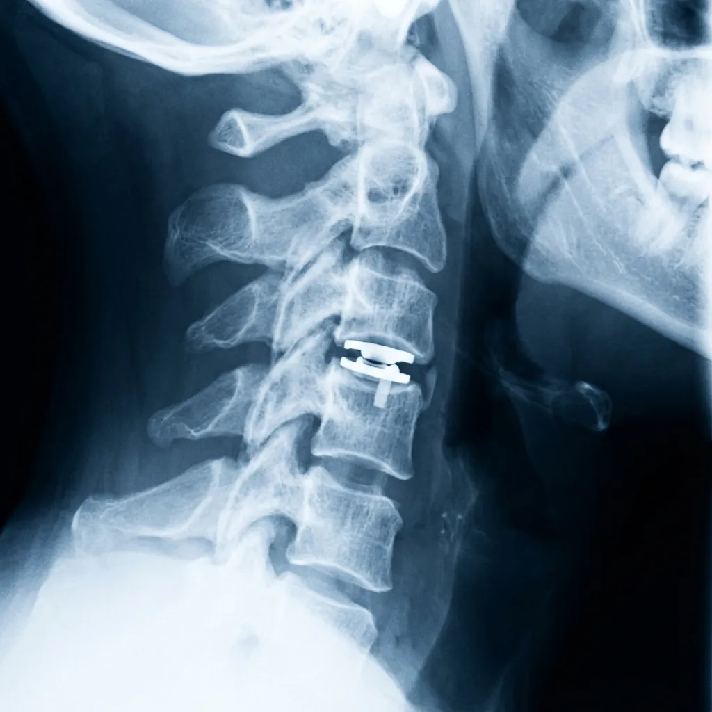
Six-Week Radiological Assessment
Imaging Protocol
Standard and dynamic views
Activity Progression at 6-Week Milestone
Rehabilitation Intensification
Goals
Interventions
Goals
Interventions
Cervical Recovery Timeline
A comprehensive overview of the cervical disc replacement recovery journey from immediate postoperative care through long-term outcomes.
Recovery Progress
Motion-preserving cervical disc replacement enables accelerated recovery with most patients returning to normal activities within 6 weeks and achieving sustained long-term outcomes through comprehensive postoperative care.
Expected Functional OutcomesRecovery Trajectories
Symptom resolution follows characteristic patterns with research demonstrating substantial improvements in pain, function, and quality of life.
Radicular Arm Pain
Most patients experience dramatic reduction immediately following surgical decompression (day 0–1).
Pain may persist if prolonged compression caused irreversible nerve damage.
Neck Pain
Gradual improvement over 3–6 months with some further improvement through 12 months.
Neck Disability Index (NDI) Progression
The gold standard for measuring cervical spine-related disability demonstrates substantial improvement. Minimal Clinically Important Difference (MCID): 10–15 points—most patients substantially exceed this threshold.
Quality of Life Improvements
Sleep Quality
Marked improvement as pain resolves; contributes to improved mood and energy
Psychological Outcomes
Depression and anxiety scores significantly reduce as pain resolves
Return to Activities
70–80% return to work by 1 year; >85% resume preoperative hobbies
Patient Satisfaction
85–95% report high satisfaction; >90% satisfied with pain improvement
CDR vs Fusion: Consistent Advantages
2025 Meta-Analysis Evidence
| Metric | CDR | Fusion | P-Value |
|---|---|---|---|
| 6-Week Return-to-Work | OR = 1.33 | — | P = 0.01 |
| 3-Month Return-to-Work | OR = 1.58 | — | P = 0.001 |
| 1-Year Return-to-Work | OR = 1.35 | — | P = 0.04 |
| Mean RTW Difference | 9.91 days earlier | — | P = 0.01 |
| Overall Reoperation Rate | 5.6% | 7.8% | |
| Adjacent Segment Disease | 1–2% | 3–5% | |
| Adjacent Segment Degeneration | 26.2% | 43.9% | P < 0.001 |
| Segmental Motion | 8–12° | 0° |
Motion Preservation Benefit: CDR maintains 8–12° of segmental motion, distributing mechanical stress more physiologically and reducing adjacent segment overload.

Physical Therapy ProtocolsEvidence-Based Guidance
Physical therapy benefit remains nuanced in current evidence, with recent high-quality studies questioning the necessity of formal supervised therapy for all patients.
Key Research Findings
Prospective RCT (NDI Outcomes)
No significant differences in Neck Disability Index between structured PT and standard care at 1-year follow-up
PROMIS Assessment Study
No significant differences in physical function scores at 6 months or 1 year between PT and non-PT groups
Patients Who Benefit Most
Patients Who May Not Require Formal PT
Shared Decision-Making Approach
Individualised recommendations
Determining which patients require formal PT should involve discussion of:
Typical Program Structure

Complications and ManagementRecognition and Intervention
Understanding potential complications enables early recognition, appropriate intervention, and realistic expectation-setting. The overall complication profile of CDR is favourable compared to fusion surgery.
Neurological Complications
New or worsening radiculopathy, myelopathy, or cranial nerve injury
Urgent imaging evaluation; implant malposition may warrant reoperation; transient symptoms typically improve with time
Dysphagia
Swallowing difficulty affecting majority immediately postoperatively
Speech pathology consultation; dietary modifications; reassurance that resolution is expected (typically 24–48 hours)
Wound Complications
Infection, haematoma, and seroma are rare but require early recognition
Prophylactic antibiotics minimise infection risk; early recognition essential for prompt intervention
Heterotopic Ossification (HO)
Most common radiological finding; varies by implant type and assessment criteria
95%+ remain asymptomatic; surveillance imaging for asymptomatic cases; intervention rarely needed
Subsidence
Implant sinking into vertebral endplates; typically clinically insignificant
Most occurs within 6–12 months then plateaus; monitoring with serial imaging
Migration
Implant displacement from initial position
Requires evaluation if significant; modern implants have very low migration rates
Adjacent Segment Degeneration (ASD)
Imaging changes by 5–10 years; symptomatic disease requiring reoperation in <5–10%
Surveillance imaging; conservative management for symptoms; adjacent level surgery rarely needed
Adjacent Segment Disease
Symptomatic degeneration requiring intervention; lower rate than fusion (3–5%)
CDR shows somewhat lower rates; motion-preserving advantage reduces adjacent segment overload
Implant Wear
Becomes a concern with very long-term follow-up; current generation shows encouraging wear characteristics
Decades of follow-up required for definitive assessment; modern implants optimised for durability
Heterotopic Ossification Risk Factors
While HO is common radiologically (30–70%), most remains clinically insignificant. Understanding risk factors helps with patient counselling.
CDR Reoperation Rate
2–5%Overall reoperation rates remain low, with lower rates than fusion approaches
Revision to Fusion
RareWhen indicated for symptomatic complications, revision to fusion surgery is typically successful

Long-Term Follow-UpSurveillance Protocols
Research demonstrates sustained benefits through long-term follow-up, with patient satisfaction and functional improvements maintained despite potential progression of heterotopic ossification or adjacent segment changes.
Clinical Follow-Up Schedule
Expected Long-Term Outcomes
Pain Relief Maintained
Neck pain and arm pain improvement sustained at long-term follow-up
Functional Improvement Sustained
Disability scores show plateau with sustained improvements
Motion Preserved
Segmental range of motion maintained in majority of cases
Satisfaction Maintained
High satisfaction rates sustained through long-term follow-up
Reoperation Rates Low
2–5% overall; lower than fusion approaches

Motion Preservation Sustainability
Long-term studies extending to 10–15 years demonstrate sustained motion preservation in most patients, though some degree of motion loss may occur due to heterotopic ossification or adjacent segment changes.
Patient Education and ExpectationsSetting Realistic Goals
Setting appropriate expectations prevents disappointment and anxiety during the recovery process. Recovery timeline varies among individuals, with some experiencing immediate relief whilst others require months for optimal recovery.
Realistic Recovery Timeline
Initial Recovery
Gradual Improvement
Significant Progress
Continued Improvement
Most Improvement Achieved
Stabilisation
Long-Term Spine Health Maintenance
Proactive measures preserve surgical benefits and optimise long-term outcomes.
Postural Awareness & Ergonomics
Physical Activity
Weight Management
Smoking Cessation
Stress Management
Continued Surveillance
Comprehensive Approach to Optimal Recovery
Successful recovery from cervical disc replacement extends far beyond the operative procedure, requiring comprehensive postoperative care addressing pain management, activity progression, rehabilitation, complication prevention, and long-term follow-up.
The principles outlined—early mobilisation, multimodal pain management, structured rehabilitation when beneficial, and realistic expectation-setting—provide the foundation for optimal outcomes.
Patients who embrace the recovery process, adhere to evidence-based protocols, maintain realistic expectations, and sustain long-term commitment to spinal health achieve excellent outcomes with symptom relief, functional restoration, and high satisfaction rates. The motion-preserving design of cervical disc replacement offers the potential for superior long-term outcomes compared to fusion surgery.