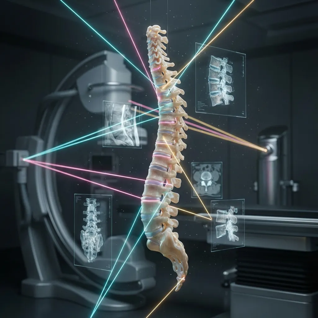
Radiological Investigations For Spinal Arthroplasty
Comprehensive imaging and diagnostic protocols that determine whether motion preservation offers superior long-term outcomes, ensuring every surgical recommendation is grounded in objective clinical evidence.
Imaging Options
Comprehensive Assessment
9 Modalities
From foundational MRI and CT through advanced SPECT/CT, Nociscan, and neurophysiological testing for complete diagnostic clarity.
Satisfaction Rate
Evidence-Based Selection
75-93%
Patients selected using comprehensive imaging criteria demonstrate excellent mid-to-long-term satisfaction with disc replacement outcomes.
Decision Framework
Clinical Correlation
Integrated
Systematic diagnostic algorithms combining anatomical imaging, functional assessment, and predictive biomechanical analysis.
Imaging as Decision-MakingArchitecture
Diagnostic imaging in spinal arthroplasty serves a fundamentally different purpose than imaging in fusion surgery. Where fusion imaging confirms degenerative pathology requiring stabilisation, arthroplasty imaging must determine whether motion preservation offers superior long-term outcomes justifying the inherent risks of implantation.
This comprehensive approach ensures radiological assessment translates directly into clinical decision-making regarding optimal treatment selection.
Whether motion preservation offers advantage justifying the inherent risks of implantation, or whether fusion's proven stability and lower operative complexity better serves the individual patient.
Three Critical Realities
- Asymptomatic individuals routinely demonstrate moderate to severe degenerative changes on imaging
- A well-established phenomenon in spine literature challenges traditional imaging interpretation
- Clinical correlation remains essential—imaging must match symptoms
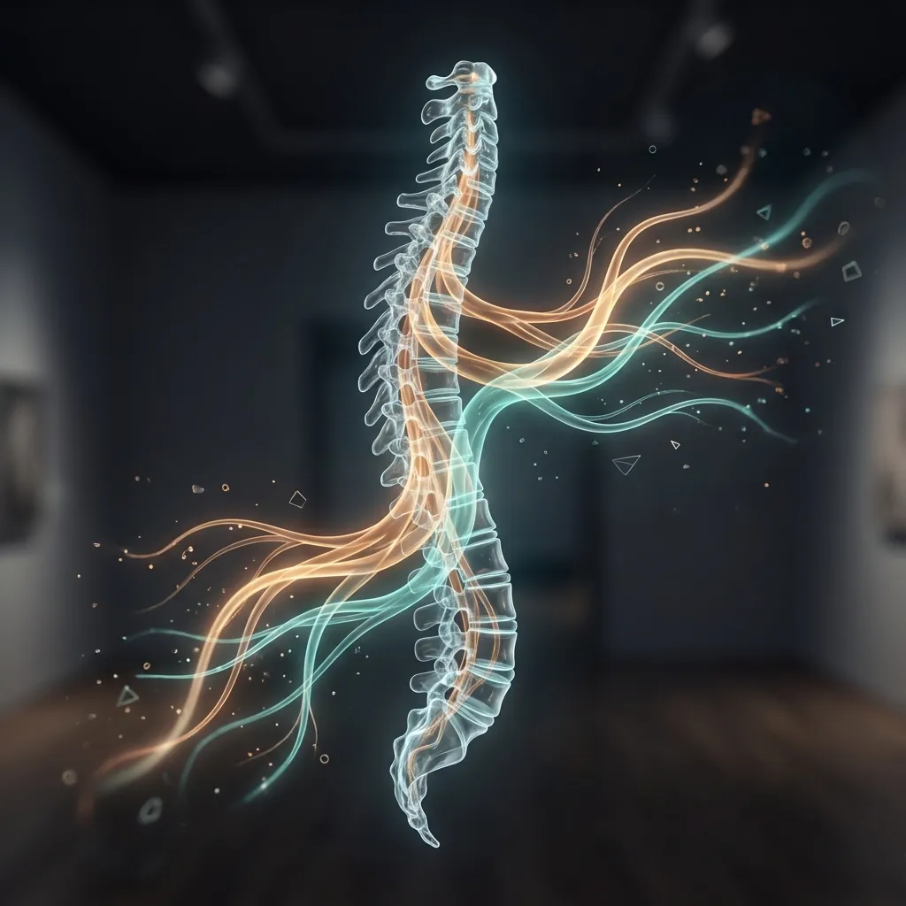
The Role of Imagingin Surgical Planning
Disc replacement candidacy requires meticulous preoperative evaluation. Imaging studies serve multiple critical functions: confirming the diagnosis, identifying contraindications, assessing adjacent segment health, documenting baseline parameters, and enabling precise surgical planning.
Diagnostic Confirmation
Imaging must confirm that patient symptoms correlate with pathological findings at the target level. This prevents unnecessary intervention for incidental imaging abnormalities—a common pitfall in modern spine practice where asymptomatic degenerative findings are nearly universal on advanced imaging.
Candidacy Assessment
Imaging identifies anatomical factors that may favour or preclude arthroplasty. Certain findings (severe osteoporosis, complete bridging osteophytes, severe facet arthropathy) substantially increase the likelihood that fusion offers more predictable outcomes than disc replacement.
Surgical Planning
Detailed imaging provides dimensional data for implant sizing, approach selection, decompression requirements, and anticipation of anatomical variants that might complicate operative access.
Baseline Documentation
Comprehensive preoperative imaging establishes reference parameters—disc height, segmental motion, adjacent segment status, heterotopic ossification, and alignment—essential for postoperative comparison and late complication detection.
The Imaging Algorithm
Sequential Assessment and Selective Advancement
Tier 1: Essential — All Patients
- Plain radiographs (neutral, flexion, extension)
- MRI of the involved spinal region
Foundation of every arthroplasty assessment
This tiered approach ensures cost-effectiveness and clinical efficiency while obtaining necessary information for informed decision-making. Not every patient requires all imaging modalities; rather, imaging is advanced sequentially based on diagnostic questions arising from initial evaluation.
Plain RadiographyStructural Assessment
Standard preoperative imaging begins with plain radiographs obtained in multiple positions to assess disc height, segmental motion, vertebral translation, spinal alignment, and osteophyte burden.
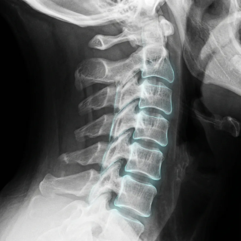
Standard Views and Protocol
Required Views
- Lateral cervical radiographs in three positions: neutral, maximum flexion, maximum extension
- Anteroposterior (AP) view
- Open-mouth odontoid view
Dynamic views require patient cooperation and genuine maximal effort—inadequate flexion or extension produces false negatives for instability assessment.
Disc Height Assessment
Progressive height loss indicates advancing degeneration with loss of nucleus pulposus hydration. Severely collapsed discs create technical challenges and carry increased perioperative complication risk.
Segmental Motion Quantification
Complete motion loss raises questions regarding whether arthroplasty can restore function. When motion loss results from osteophyte impingement, arthroplasty with osteophyte removal often successfully restores motion.
Vertebral Translation Assessment
Anterior-posterior translation of vertebral bodies measured on sagittal radiographs identifies spondylolisthesis or other instability patterns. Significant translation (Grade 2 or greater) or dynamic instability suggests fusion may be more appropriate than disc replacement.
| Grade | Description | Translation |
|---|---|---|
| Grade 0 | No slip | 0% |
| Grade 1 | Minimal slip | <25% |
| Grade 2 | Moderate slip | 25-50% |
| Grade 3 | Significant slip | 50-75% |
| Grade 4 | Severe slip | >75% |
Limitations of Plain Radiography
Plain radiographs provide limited information regarding bone density, facet joint detail, soft tissue anatomy, and neural compression severity. They cannot assess disc hydration status (essential for disc replacement candidacy). Metal artefacts from existing hardware severely degrade image quality. For patients considering arthroplasty, plain radiographs alone are insufficient for definitive surgical decision-making.
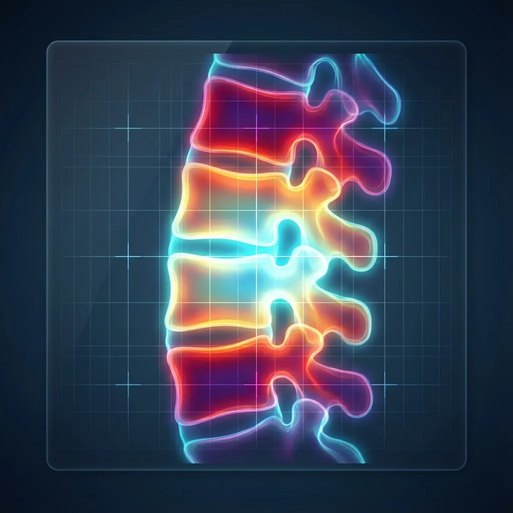
Magnetic Resonance ImagingDisc Assessment
MRI creates detailed cross-sectional images through manipulation of hydrogen nuclei in a powerful magnetic field without ionising radiation. It is the gold standard for disc and soft tissue assessment.
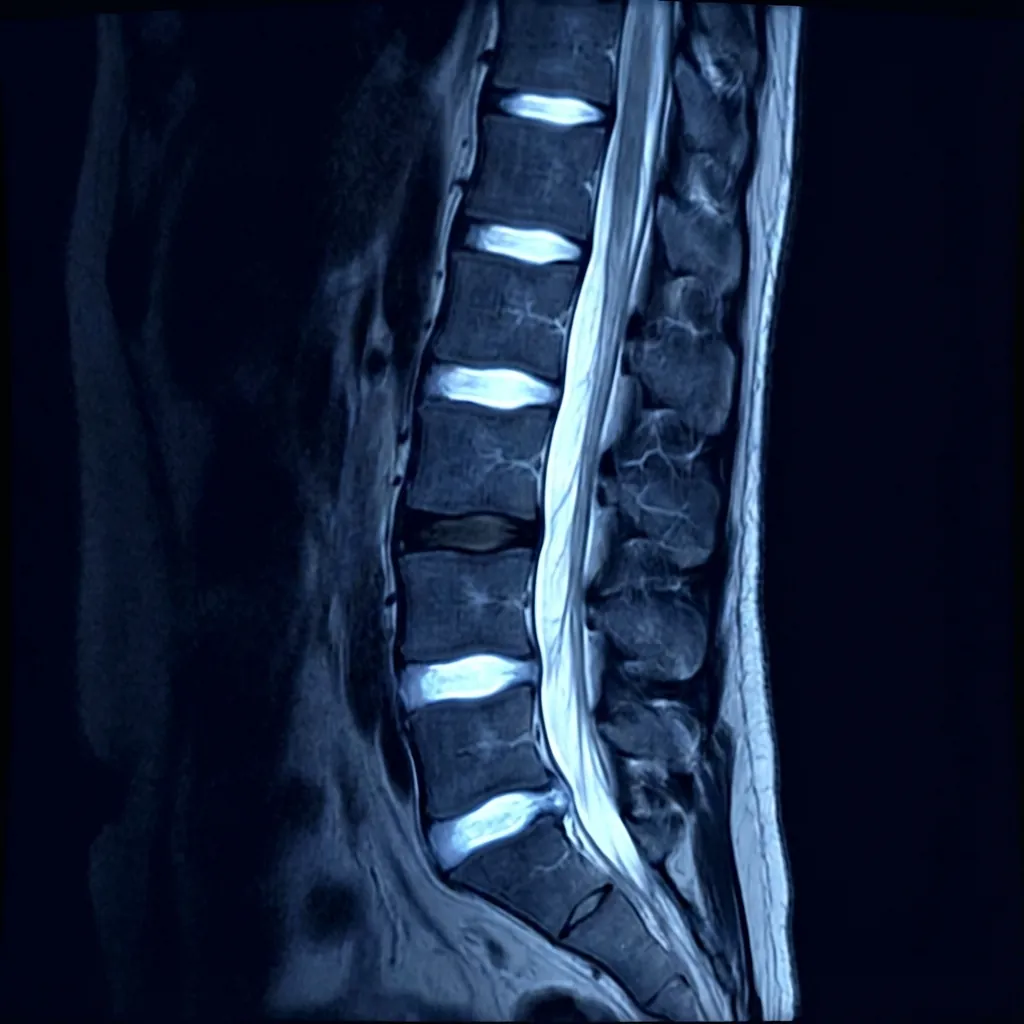
Standard Sequences and Clinical Applications
| Sequence | Primary Purpose | Arthroplasty Value |
|---|---|---|
| T2-Weighted Sagittal | Disc hydration assessment, spinal cord signal, overall alignment | Pfirrmann grading, myelopathy detection, overall severity |
| T2-Weighted Axial | Neural foramina patency, lateral recess stenosis, disc morphology | Disc-osteophyte complexes, foraminal stenosis, facet detail |
| T1-Weighted Sagittal | Anatomical detail, marrow signal, endplate integrity | Modic changes, vertebral body height, fat content |
| STIR/Fat-Saturated T2 | Bone marrow oedema, active inflammation, cord changes | Active inflammatory processes, myelomalacia |
| Gradient Echo (GRE) | Osteophyte detection, calcification sensitivity | OPLL detection, posterior element ossification |
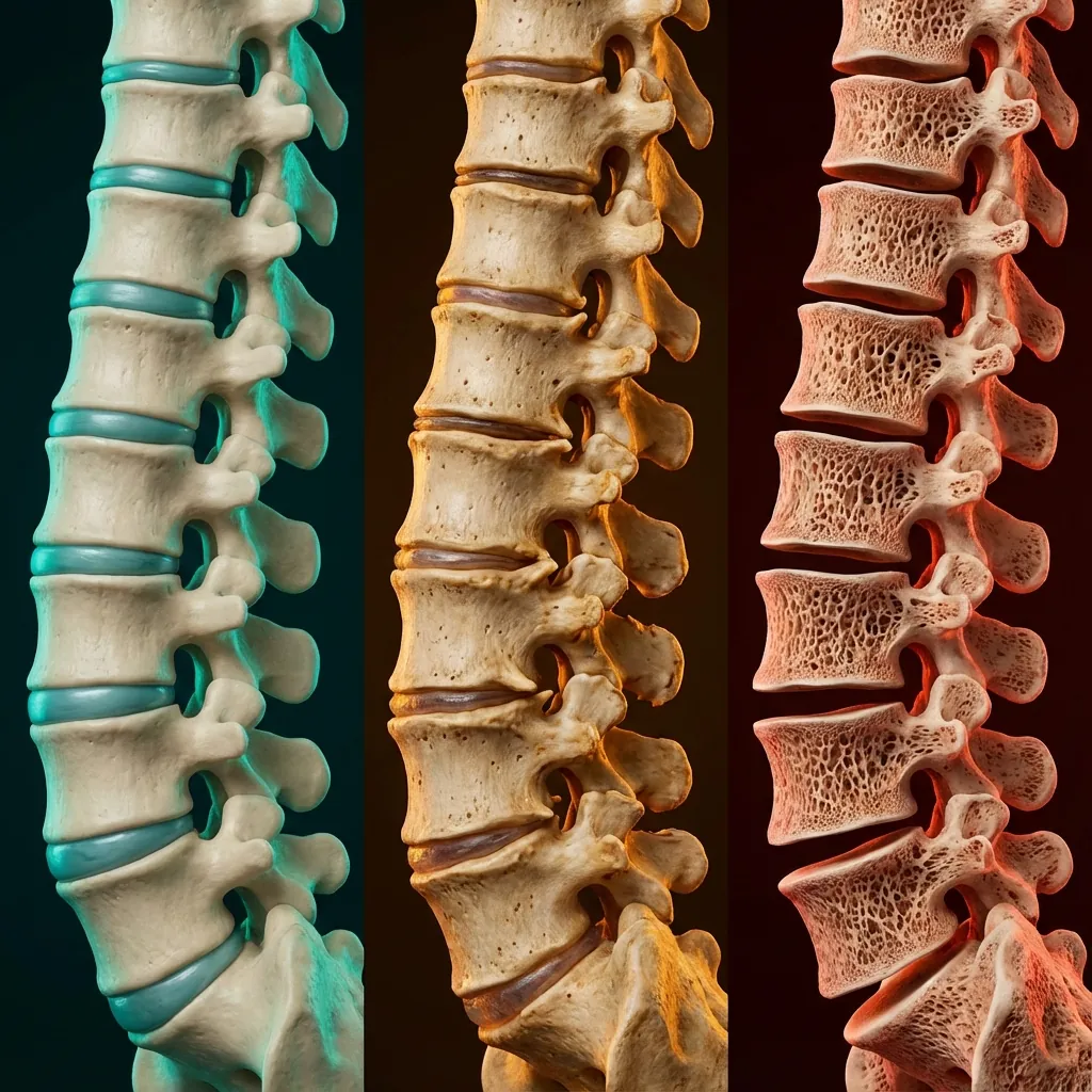
Pfirrmann Grading System
The modified Pfirrmann scale quantifies disc degeneration progression with established inter-observer reliability. Grades 1-4 generally permit disc replacement; Grade 5+ creates technical challenges.
Grade 3: Moderate Degeneration
Suitable for ArthroplastyNoticeably darker
Less distinct
Slight loss
Modic Endplate Changes
Approximately 50-80% of symptomatic discs demonstrate Modic changes; their presence suggests pain may be discogenic and potentially responsive to motion-preserving treatment.
Dark
Bright
Pathology: Inflammatory response, hypervascularity
Clinical Significance: Suggests inflammatory processes that often improve with disc restoration and motion preservation
Facet Joint Assessment
Normal joint space, no osteophytes
Minimal joint space narrowing, small osteophytes
Moderate joint space narrowing, moderate osteophytes, possible capsular hypertrophy
Severe joint space narrowing, large osteophytes, severe degenerative changes
Severe facet arthropathy (Grade 3 changes) combined with significant facet-mediated symptoms may suggest fusion as more appropriate than disc replacement, which preserves motion and may increase facet loading.
MRI Advantages
- Gold standard for disc hydration status and Pfirrmann grading
- Assessing spinal cord compression and myelopathic changes
- Identifying ligamentous ossification patterns
- Evaluating nerve root compression
- Detecting bone marrow pathology (Modic changes)
- No ionising radiation—appropriate for serial imaging
MRI Limitations
- Limited bone cortical detail compared to CT
- Dense osteophytes may be underestimated
- Metal artefacts from existing hardware degrade quality
- Contraindicated with pacemakers or ferromagnetic implants
- Long acquisition time (30-45 minutes)
- Higher cost than CT for bone-detail assessment
Computed TomographyThree-Dimensional Assessment
CT creates cross-sectional bone images through differential X-ray absorption, offering unmatched spatial resolution for bone detail. Unlike MRI's superior soft tissue characterisation, CT excels at demonstrating precise bone anatomy essential for surgical planning.
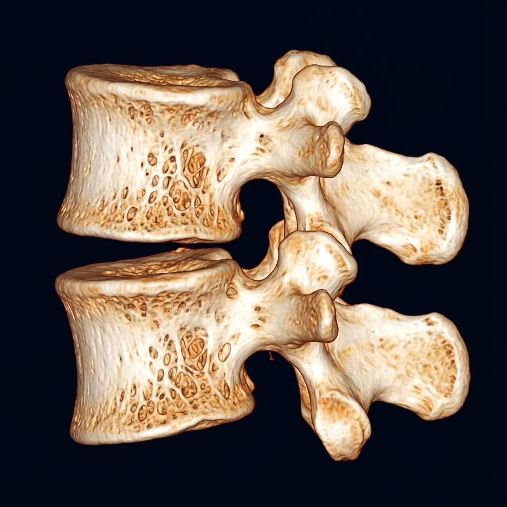
Bone Quality Assessment
Cortical thickness, trabecular pattern, and overall bone stock evaluation for implant subsidence risk prediction
Osteophyte Mapping
Precise localisation for surgical resection planning, bridging osteophyte identification, OPLL assessment
Vertebral Dimensions
Accurate endplate measurements for implant sizing ensuring optimal device seating and coverage
Fusion Assessment
Prior fusion mass integrity evaluation if revision surgery considered
Critical Distinction: OPLL vs Osteophytosis
Essential differentiation for surgical planning
OPLL (Ossification of PLL)
Continuous ossification of the posterior longitudinal ligament creating canal stenosis. Appears on CT as continuous ossified band posterior to vertebral bodies.
⚠ Anterior approach disc replacement is relatively contraindicated at levels with significant OPLL
Standard Osteophytes
Focal bony projections arising from uncovertebral or facet joints. Can be surgically removed during arthroplasty to restore motion.
✓ Osteophyte removal during arthroplasty often successfully restores motion
Osteophyte Characterisation
Location: Anterior vertebral margin
Impact: May impinge on vascular structures during anterior approach; motion restriction during flexion
Management: Small osteophytes typically do not affect outcomes; large osteophytes may require osteotomy
Quantitative Measurements
Vertebral Body Dimensions
Minimum for secure implant fixation
Ensures adequate implant seating
Disc Space & Foramen
Technical difficulties if collapsed >50%
<5mm indicates significant stenosis
Spinal Canal
Significant compression
Partial compression
Adequate neural space
CT Protocol Specifications
- Thin-slice (≤1mm) helical acquisition
- Sagittal, coronal, and axial multiplanar reformats
- 3D volume rendering for complex anatomy
- CT myelography if MRI contraindicated
Technical Considerations
- Metal Artefact: CT produces substantially less artefact than MRI with existing hardware
- Radiation Dose: Approximately 1-3 mSv—clinically justified for surgical planning
- Bone Quality: Visual assessment can identify osteoporotic changes before DEXA manifests
DEXA ScanningImplant Fixation Prediction
Successful disc replacement requires secure implant fixation into vertebral bone. When bone is weak (osteoporotic), implants may subside into vertebral bodies, compromising biomechanics and frequently necessitating revision surgery.
Mechanical Load Considerations
Lumbar disc replacement has more stringent bone quality requirements than cervical replacement. Cervical spine supports ~4-5 kg head weight; lumbar spine supports entire trunk weight plus muscle-generated compressive forces exceeding 1000 kg during heavy lifting. These massive loads necessitate robust bone fixation for lumbar devices.
T-Score Classification and Arthroplasty Candidacy
| T-Score | Classification | Bone Status | Candidacy |
|---|---|---|---|
| > -1.0 | Normal | Healthy bone density | Excellent |
| -1.0 to -2.5 | Osteopenia | Low bone density | Good |
| -2.5 to -3.5 | Moderate Osteoporosis | Significantly reduced | Questionable |
| -3.5 to -4.0 | Severe Osteoporosis | Severely reduced | Marginal |
| < -4.0 | Very Severe Osteoporosis | Extremely reduced | Contraindicated |
Moderate Osteoporosis (T-score -2.5 to -3.5)
Grey zone requiring individualised decision-making. Subsidence occurs in approximately 10-15% of cases but often stabilises without clinical consequence.
Moderate Osteoporosis: The Grey Zone
Patients with moderate osteoporosis (T-score -2.5 to -3.5) require individualised decision-making. Key considerations include:
Age & Life Expectancy
Younger patients benefit more from motion preservation
Activity Level
Higher activity increases implant loading stress
Comorbidities
General health status affects healing
Patient Preference
Understanding of risks is essential
Bone-Strengthening Management
When moderate osteoporosis is discovered in an otherwise excellent candidate
Bisphosphonates
Alendronate, risedronateInhibit bone resorption
Denosumab
RANKL antibodyPotent antiresorptive
Teriparatide
Recombinant PTHStimulates bone formation
Vitamin D & Calcium
SupplementationFoundation substrate
Treatment Protocol
Repeat DEXA scanning 3-6 months after initiating bone-strengthening therapy. T-score typically improves 0.3-0.8 points with optimal treatment compliance. If adequate improvement achieved (T-score greater than -2.5), disc replacement becomes appropriate consideration. Postoperatively, bone-strengthening medications continued to maintain bone density and minimise long-term subsidence risk.
SPECT/CTFacet Assessment
Single Photon Emission Computed Tomography combined with CT (SPECT/CT) provides functional information about metabolic activity overlaid on anatomical images—distinguishing actively inflamed or degenerating structures from stable chronic changes.
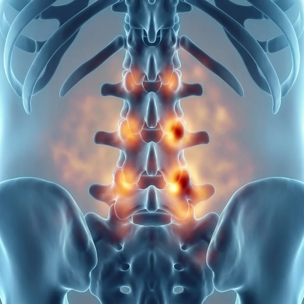
Technical Principles
SPECT uses a bone-seeking radioactive tracer (technetium-99m labelled bisphosphonate) that concentrates in areas of increased bone metabolism. When fused with CT images, the functional data is precisely localised to specific anatomical structures—enabling correlation of metabolic activity with degenerative changes.
Clinical Application: Hot vs Cold Facets
Metabolically Active ("Hot") Facets
- Increased radiotracer uptake on SPECT
- Active bone remodelling or inflammation
- May correlate with clinical facet pain
Patient Selection Indications
- Ambiguous clinical presentation with multilevel degenerative changes
- Positive response to diagnostic facet blocks requiring confirmation
- Distinguishing discogenic from facetogenic pain contribution
- Identifying specific painful levels in complex multilevel disease
- Confirming or excluding inflammatory arthropathy
Limitations & Considerations
- Limited specificity—uptake indicates bone turnover but not necessarily pain
- Additional radiation exposure beyond standard CT
- Limited availability at all centres
- Insurance coverage may be inconsistent
- Interpretation requires experienced nuclear medicine physician
Nociscan MRIDisc Spectroscopy
Nociscan represents a paradigm shift in disc assessment—using MRI spectroscopy to identify biochemical markers of painful disc degeneration without injection or provocation. It provides objective, quantitative data about disc pathology.
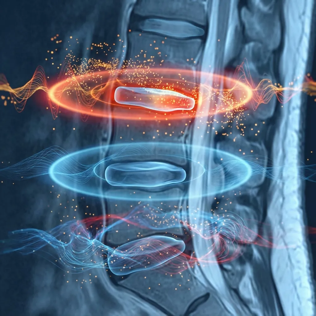
The Nociscore Concept
Nociscan generates a Nociscore for each disc—a quantitative measure of biochemical markers associated with painful degeneration. High Nociscore levels correlate with discogenic pain sources; low scores suggest the disc is unlikely to be a significant symptom generator, regardless of morphological abnormalities on conventional MRI.
Biochemical Markers Assessed
Lactate
Elevated lactate suggests anaerobic metabolism associated with disc degeneration and nerve ingrowth
Correlates with inflammatory pain pathways
Alanine
Amino acid marker elevated in degenerative disc tissue
Indicates cellular stress and altered metabolism
Proteoglycan Degradation
Breakdown products of disc matrix components
Direct measure of structural degeneration
Collagen Fragments
Markers of collagen breakdown in the nucleus and annulus
Reflects structural disc failure
Advantages Over Traditional Discography
No Injection Required
Unlike discography, purely non-invasive imaging protocol
Objective Measurement
Quantitative biochemical data rather than subjective pain response
No Procedural Risks
Eliminates disc puncture, infection, or acceleration of degeneration
Reproducible Results
Less operator-dependent than provocative testing
Multiple Levels
All discs assessed simultaneously in single examination
FDA Cleared
Regulatory approval for clinical use in the United States
Clinical Application
Nociscan is particularly valuable when conventional imaging shows multilevel disc degeneration and the specific painful level is uncertain. It can:
- Identify which morphologically abnormal disc(s) are symptomatic
- Guide surgical level selection in multilevel disease
- Reduce need for invasive provocative discography
- Support conservative management when Nociscore is low
Current Considerations
- Relatively new technology with evolving clinical evidence
- Not universally available at all imaging centres
- Requires compatible MRI scanner and software
- Insurance coverage varies by region and payer
- Best used as part of comprehensive assessment, not sole determinant
Vascular ImagingAnterior Approach Surgery
Lumbar disc replacement typically requires anterior approach, necessitating mobilisation of major abdominal vessels. Preoperative vascular imaging identifies anatomical variants and pathology that might complicate or contraindicate anterior access.
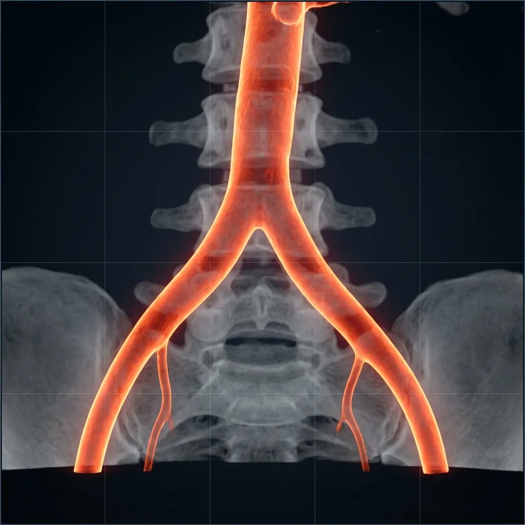
Anatomical Variants (15% of Population)
Approximately 15% of patients have vascular anatomy that complicates or precludes standard anterior approach. These variants include high aortic bifurcation, low-lying iliac veins, aberrant vessels, or significant atherosclerotic disease. Unrecognised vascular anomalies are a preventable cause of major intraoperative haemorrhage.
Systematic Vascular Assessment
Aorta
Calcification, aneurysm, atherosclerotic disease
Inferior Vena Cava
Position, compression, thrombosis
Common Iliac Arteries
Bifurcation level relative to L4-L5, calibre, atherosclerosis
Common Iliac Veins
Position, patency, confluence level
Left Common Iliac Vein
Compression between aortic bifurcation and spine (May-Thurner anatomy)
Sacral Vessels
Middle sacral artery and vein position
CT Angiography (CTA)
Gold StandardAdvantages
- Precise vascular anatomy with high spatial resolution
- Comprehensive assessment of calcification and atherosclerosis
- Accurate measurement of vessel-to-spine distances
- 3D reconstruction for surgical planning
- Rapid acquisition (seconds)
Limitations
- Ionising radiation exposure
- Contrast-related risks (allergy, nephrotoxicity)
- May overestimate calcification extent
MR Angiography (MRA)
AlternativeAdvantages
- No ionising radiation
- Gadolinium contrast safer for renal patients
- Can be combined with routine MRI
Limitations
- Lower spatial resolution than CTA
- Longer acquisition time
- Less accurate calcification assessment
- Contraindicated with some implants
Ultrasound Screening
AdjunctAdvantages
- Non-invasive, no radiation
- Real-time assessment
- Identifies DVT or significant stenosis
- Low cost
Limitations
- Operator-dependent
- Limited visualisation of deep structures
- Cannot provide detailed surgical planning
- Not suitable as sole preoperative assessment
Diagnostic InjectionsPain Source Identification
When imaging demonstrates multilevel pathology, diagnostic injections help identify the specific pain-generating structures. For arthroplasty candidates, distinguishing discogenic from facetogenic pain is critical—facet-predominant pain may respond better to fusion.
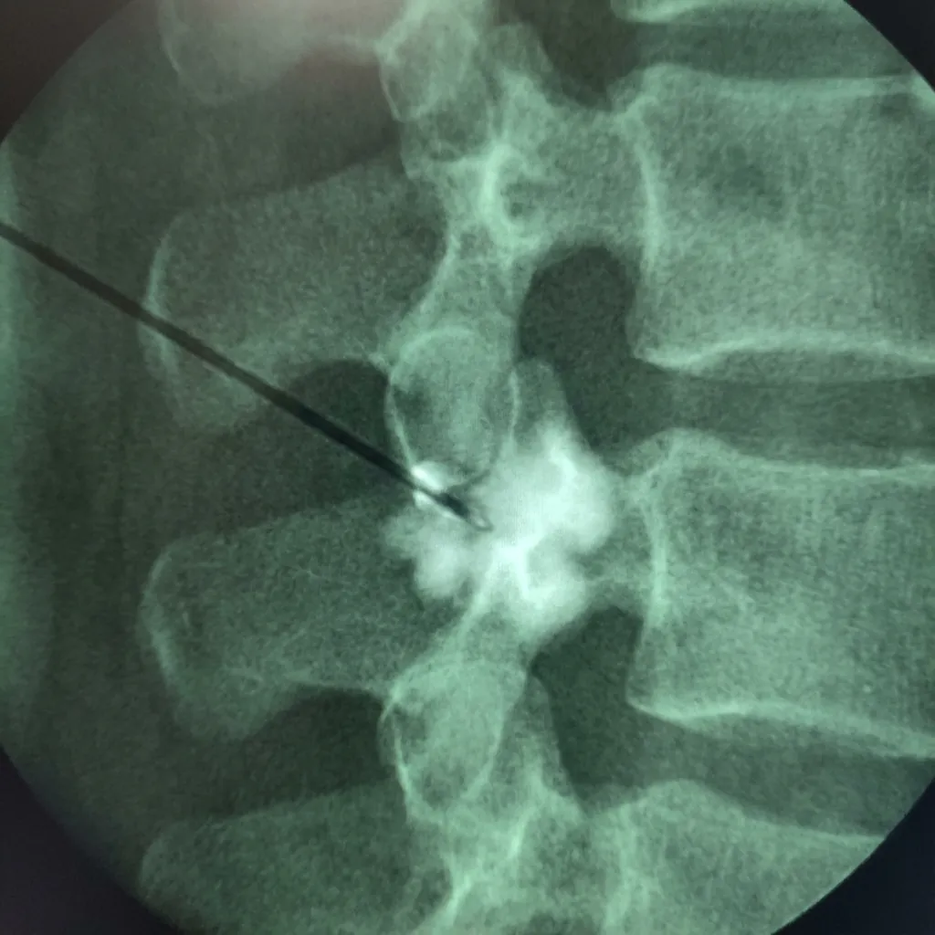
Critical Role in Treatment Selection
In patients with concurrent degenerative disc disease and facet arthropathy (common multilevel pathology), diagnostic injections help identify which pathology generates symptoms. If facets are primary pain generators, disc replacement may perpetuate or worsen facet pain by maintaining facet loading. Fusion, which eliminates facet motion, may provide better outcomes for facet-predominant symptoms.
Injection Approaches
Medial Branch Block (MBB)
Anaesthetises sensory nerves before entering joint; if pain relief occurs, confirms facet as pain generator
- Standardised and reproducible technique
- Can be repeated for diagnostic confirmation (double block)
- Enables subsequent radiofrequency ablation if positive
- Lower infection risk than intra-articular injection
- May not fully anaesthetise joint capsule inflammatory component
- False positives from anaesthetic spread to adjacent structures
- Temporary relief only; requires follow-up procedure for treatment
Comparison: Medial Branch Block vs Intra-Articular Injection
| Feature | Medial Branch Block | Intra-Articular |
|---|---|---|
| Primary Target | Medial branch nerves | Facet joint capsule |
| Diagnostic Accuracy | 75-85% | 70-80% |
| Technical Difficulty | Moderate | Higher |
| Infection Risk | Lower | Higher |
| Follow-on Treatment | RF ablation eligible | Repeat injection or ablation |
| False Positive Rate | 25-40% (single block) | 30-45% |
Double Block Protocol
Gold standard for reducing false positive rates
First Block
Initial medial branch block performed with local anaesthetic alone
Pain Assessment
Patient completes pain diary for 6-8 hours post-injection
Interval Period
Minimum 2-week interval before second block
Second Block
Repeat block with different anaesthetic (lidocaine if bupivacaine used first, or vice versa)
Concordant Response
Positive diagnosis requires ≥75-80% relief on both occasions with appropriate duration differences
Single block false positive rate is 25-40%. The double-block protocol reduces this to approximately 10-15%, substantially improving diagnostic specificity and subsequent treatment selection accuracy.
Provocative DiscographyDisc Pain Identification
Provocative discography involves controlled pressurisation of intervertebral discs to identify pain-generating levels. When a disc is pressurised and reproduces the patient's typical pain pattern, it suggests that disc is a symptom source.
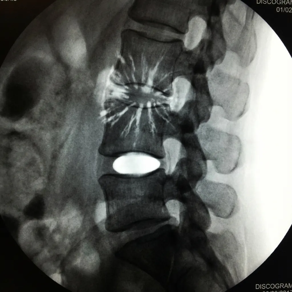
Contemporary Role
Once the gold standard for identifying painful discs, discography's role has evolved. Modern non-invasive alternatives (particularly Nociscan MRI spectroscopy) increasingly provide objective diagnostic information without procedural risks. However, discography remains valuable when non-invasive testing is inconclusive or unavailable.
Dallas Discogram Description (Modified)
Grade 3: Outer Annular Tear
Contrast extends to outer third of annulus
Indications
- Multilevel degenerative changes where single symptomatic level requires identification
- Discordance between clinical presentation and imaging findings
- Previous failed spine surgery requiring assessment of adjacent levels
- When Nociscan is unavailable or inconclusive
- Confirmation of suspected internal disc disruption
Contraindications
- Active infection
- Bleeding diathesis
- Known disc space infection
- Patient unable to provide reliable feedback
- Previous adverse reaction to contrast
- Psychogenic pain disorder diagnosis
- Workers compensation claims (reduced reliability)
- Severe anxiety or pain behaviour
Discography vs Nociscan Comparison
| Feature | Provocative Discography | Nociscan MRI |
|---|---|---|
| Invasiveness | Invasive (needle puncture) | Non-invasive (MRI only) |
| Pain Provocation | Required for diagnosis | Not required |
| Objective Data | Morphology + subjective pain | Quantitative biochemical markers |
| Infection Risk | Present (0.1-0.25%) | None |
| Disc Acceleration Risk | Theoretical concern | None |
| Specificity | Variable (depends on technique) | Improving with validation studies |
| Availability | Widely available | Limited centres |
Clinical Recommendation: When Nociscan is available and shows concordant results with clinical presentation, provocative discography may be unnecessary. However, when Nociscan is inconclusive, unavailable, or when results conflict with clinical impression, discography remains a valuable diagnostic tool.
EMG & NCS StudiesNerve Function Assessment
Electrodiagnostic studies assess the physiological function of peripheral nerves rather than anatomical structure. This provides complementary information to imaging, distinguishing active from chronic nerve damage and correlating structural findings with functional consequences.
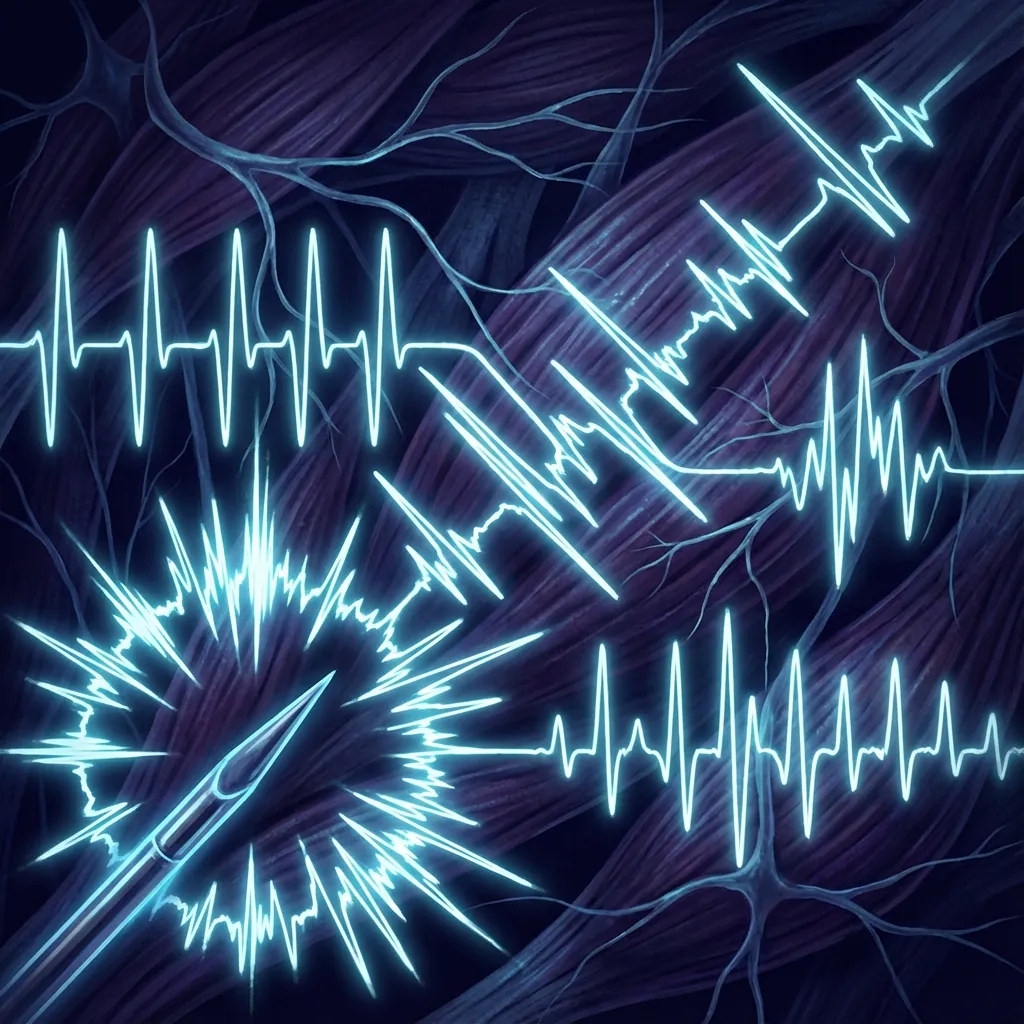
Clinical Value in Arthroplasty Assessment
EMG/NCS helps identify patients who will benefit from decompression during arthroplasty and predicts functional recovery potential. Active denervation on EMG combined with imaging evidence of compression provides strong evidence that surgical decompression will provide meaningful neurological improvement.
Study Components
Insertional Activity
Brief electrical activity when needle enters muscle; prolonged activity suggests denervation
Spontaneous Activity
Fibrillation potentials and positive sharp waves indicate active denervation
Motor Unit Action Potentials
Size, shape, and recruitment patterns indicate reinnervation or chronic changes
Recruitment Pattern
Reduced recruitment indicates nerve root pathology affecting that myotome
Diagnostic Accuracy by Radiculopathy Severity
| Clinical Condition | Sensitivity | Specificity |
|---|---|---|
| Definite Radiculopathy | 85-95% | 80-90% |
| Moderate Radiculopathy | 70-80% | 75-85% |
| Mild/Subclinical | 40-60% | 70-80% |
| Chronic Stable | 60-70% | 70-80% |
EMG sensitivity increases with radiculopathy severity. Mild or subclinical radiculopathy may produce normal EMG findings despite genuine nerve root involvement.
EMG/NCS vs Imaging: Complementary Roles
| Feature | EMG/NCS | MRI/CT |
|---|---|---|
| Primary Assessment | Nerve function/physiology | Anatomical structure |
| Temporal Information | Acute vs chronic changes | Snapshot at single time |
| Active Denervation | Directly detected | Inferred from compression |
| Asymptomatic Findings | Less common | Very common |
| False Positives | Lower rate | Higher rate (degenerative changes) |
| Operator Dependence | High (examiner skill) | Moderate (interpretation) |
Predictive Value for Surgical Outcomes
Active denervation + imaging compression
Excellent surgical outcomes expected
Chronic changes only + imaging compression
Moderate improvement expected
Normal EMG + imaging compression
Consider conservative management longer
Denervation without imaging correlation
Search for alternative pathology
Limitations: EMG requires 2-3 weeks after nerve injury to show abnormalities. Normal EMG does not exclude radiculopathy—it may simply be too early, too mild, or compensated. EMG is also highly operator-dependent, requiring experienced neurophysiologists for reliable interpretation.
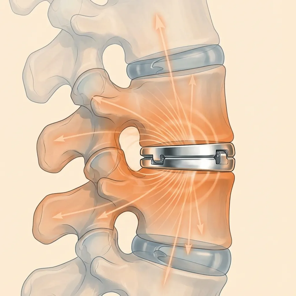
Biomechanical IntegrationOutcome Prediction
Understanding how preoperative imaging predicts long-term biomechanical outcomes enables more accurate patient counselling and treatment selection. The goal of disc replacement is not merely to remove a painful disc but to restore near-normal segmental biomechanics.
Motion Preservation Clinical Evidence
| Study/Source | Key Finding | Clinical Significance |
|---|---|---|
| FDA IDE Studies (5-7 year) | 75-85% maintained motion at treated level | Majority of devices maintain intended function |
| Long-term Registry Data | Mean ROM 6-10° at 10 years | Physiological motion preserved over time |
| Adjacent Segment Analysis | 2-5% vs 8-12% symptomatic ASD (arthroplasty vs fusion) | Reduced adjacent segment disease burden |
| Revision Rate Comparison | 5-8% vs 10-15% at 10 years | Lower long-term revision rates with motion preservation |
Favourable Predictors
- Preserved disc height (Pfirrmann ≤4)
- Preserved segmental motion (>5° ROM)
- Normal to osteopenic bone (T-score > -2.5)
- Minimal facet arthropathy (Grade 0-1)
- Contained disc pathology without severe deformity
Unfavourable Predictors
- Severe disc collapse (<2mm height)
- Rigid segment (<3° ROM)
- Osteoporotic bone (T-score < -2.5)
- Severe facet arthropathy (Grade 3)
- Bridging osteophytes or OPLL
- Instability or significant deformity
Heterotopic Ossification Classification
Long-term imaging surveillance monitors for heterotopic ossification (HO)—ectopic bone formation around the prosthesis that may eventually limit motion. Understanding HO grades helps predict long-term functional outcomes.
No heterotopic ossification
40-50%
Small islands of ossification
25-30%
Possible bridging without complete ankylosis
15-20%
Bridging ossification limiting motion
5-10%
Complete fusion (spontaneous ankylosis)
2-5%
Intradiscal Pressure Dynamics
Finite element studies demonstrate that disc replacement reduces abnormal stress transmission to adjacent segments compared to fusion. By maintaining near-physiological motion and load distribution, arthroplasty theoretically protects adjacent discs from accelerated degeneration—the primary long-term advantage over fusion surgery.
Clinical DecisionAlgorithms
Synthesising the wealth of imaging and diagnostic data requires a systematic approach to avoid cognitive errors and ensure all relevant factors are considered. This structured framework guides clinicians through the decision-making process.

Systematic 7-Step Interpretation Algorithm
Create Problem List
Document all clinical and imaging abnormalities
Imaging Red Flags
| Finding | Imaging Appearance | Required Action |
|---|---|---|
| Active Infection | Disc space narrowing with endplate destruction | Contraindicated for arthroplasty—treat infection first |
| Malignancy | Lytic or blastic lesions, pedicle destruction | Oncological evaluation before any spine surgery |
| Significant Instability | >4mm translation or >15° angular motion | Fusion typically preferred for stabilisation |
| Severe Osteoporosis | T-score <-3.5, cortical thinning on CT | Fusion more predictable; consider bone strengthening first |
| OPLL | Continuous ossification posterior to vertebral bodies | Relative contraindication at affected levels |
| Severe Facet Arthropathy | Grade 3 changes with positive facet blocks | Consider fusion to eliminate facet loading |
Disc Replacement vs Fusion: Decision Criteria
Anatomy
- Preserved disc height
- Minimal facet changes
- Normal alignment
- Adequate bone quality
- Collapsed disc
- Severe facet arthropathy
- Deformity/instability
- Osteoporosis
Pain Source
- Discogenic predominant
- Nociscan positive
- Negative facet blocks
- Facetogenic significant
- Instability-related
- Positive facet blocks
Patient Factors
- Younger/active patient
- Single or two-level disease
- No prior fusion at level
- Older patient with limited activity goals
- Multilevel disease
- Prior failed arthroplasty
Postoperative ImagingComplication Surveillance
Systematic postoperative imaging surveillance documents baseline device performance, detects early complications, and monitors long-term outcomes including motion preservation and adjacent segment health.
Baseline Postoperative Radiographs
Immediate postoperative imaging establishes essential reference parameters for all subsequent surveillance:
Device Position
Centred, proper depth, appropriate lordosis/kyphosis
Endplate Coverage
Adequate footprint size, no overhang or underhang
Disc Height
Restoration to normal or near-normal height
Alignment
Segmental and global alignment preserved or improved
Adjacent Segments
Baseline reference for future comparison
Serial Radiographic Surveillance
| Timepoint | Focus | Imaging Protocol |
|---|---|---|
| 6 Weeks | Wound healing, device stability, early subsidence detection | Standing AP/lateral |
| 3 Months | Early motion, alignment maintenance, symptomatic correlation | Standing neutral + flexion/extension |
| 6 Months | Motion quantification, heterotopic ossification screening | Standing neutral + flexion/extension |
| 12 Months | Definitive motion assessment, HO grading, subsidence evaluation | Complete series |
| 2 Years | Long-term stability, adjacent segment surveillance | Standing neutral + flexion/extension |
| 5+ Years | Late complications, wear, adjacent segment disease | Annual or as clinically indicated |
MRI After Disc Replacement
Moderate artefact, interpretable
Significant artefact, limited interpretation
Minimal artefact
Always verify specific device MRI compatibility and field strength limits before scanning.
CT for Late Complications
CT imaging is preferred when evaluating:
- Suspected device subsidence or migration
- Heterotopic ossification requiring surgical revision
- Periprosthetic osteolysis or wear-related bone loss
- Adjacent segment structural pathology
- Planning revision surgery
Case StudiesReal-World Application
These representative cases illustrate how comprehensive imaging assessment guides treatment selection. Each case demonstrates the integration of multiple diagnostic modalities into coherent clinical decision-making.
42-year-old male, active professional, non-smoker
Right arm radiculopathy and neck pain for 8 months
Failed conservative management including physiotherapy and epidural injection. No prior surgery.
- C6 dermatomal sensory changes
- Biceps weakness (4+/5)
- Preserved neck motion
- Positive Spurling test right
MRI
- • C5-6 disc protrusion with right foraminal stenosis
- • Pfirrmann Grade 3 disc
- • Modic Type 1 changes at C5-6
- • Normal adjacent segments
CT
- • Minimal uncovertebral osteophytes
- • Normal bone quality (no cortical thinning)
- • No OPLL
X-ray
- • Preserved disc height (5mm)
- • Normal segmental motion (10°)
- • Neutral cervical alignment
C6 radiculopathy secondary to single-level disc herniation at C5-6
Cervical disc replacement at C5-6
Key Decision Rationale
Conclusion
Comprehensive radiological investigation remains fundamental to appropriate patient selection for spinal arthroplasty. The integration of anatomical imaging, functional assessment, diagnostic procedures, and clinical correlation ensures that treatment recommendations are individualised and evidence-based.
Integrated Diagnostic Approach
The imaging assessment begins with plain radiographs and MRI as foundational studies, progresses through CT and DEXA for surgical planning and bone quality assessment, and employs selective advanced investigations (SPECT/CT, Nociscan, diagnostic injections, EMG) when clinical questions remain unanswered.
This systematic approach ensures that disc replacement candidates are selected based on objective imaging criteria rather than subjective clinical impression alone. The result is appropriate patient selection that optimises long-term outcomes while minimising the risk of treatment-related complications.
When comprehensive imaging assessment identifies features favouring fusion over disc replacement, this guidance prevents suboptimal outcomes from inappropriate motion-preservation attempts. Equally importantly, when imaging demonstrates excellent arthroplasty candidacy, patients can be confidently offered motion-preserving surgery with realistic expectations for long-term success.
Key Points Summary
- Plain radiographs provide foundational assessment of disc height, segmental motion, and alignment—essential baseline for all arthroplasty candidates
- MRI remains the gold standard for disc hydration assessment, Pfirrmann grading, and soft tissue characterisation
- CT provides superior bone detail for surgical planning, osteophyte mapping, and bone quality visual assessment
- DEXA scanning is essential before lumbar arthroplasty; T-score determines candidacy threshold
- SPECT/CT offers functional information distinguishing metabolically active from stable pathology